What is meibography?
Diagnostics6 September 20264 min read
Meibography is one of the key examinations in dry eye disease: it shows non-invasively how much functional gland tissue remains within the lid margins.
Medically reviewed by the ophthalmic team of Lux Augenzentrum, Zurich· Last medically reviewed: 2026-09-15
How the examination works
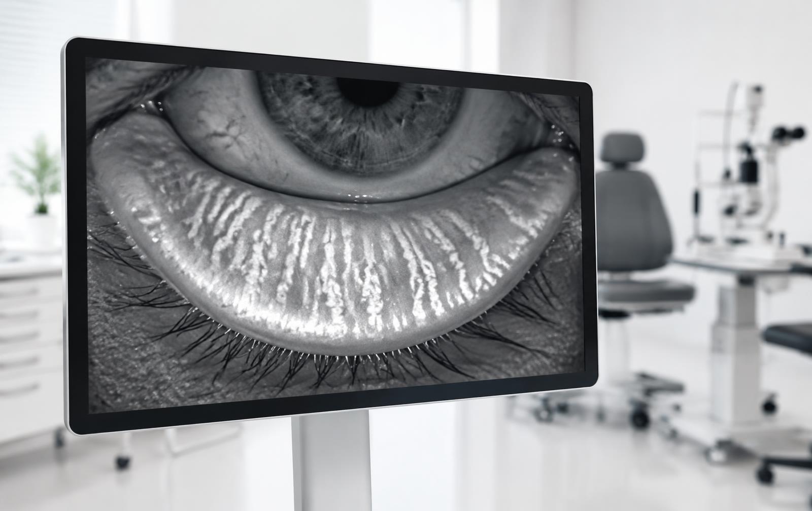
The lid is gently everted and imaged with infrared light. The capture takes only seconds, needs no drops and is not painful, although everting the lid can feel briefly unfamiliar.
In the image the glands appear as elongated bright structures. Missing, shortened or irregular areas suggest gland loss (drop-out).
What the image does and does not show
Meibography shows structure, not function. It answers how much gland tissue is present, not automatically how much lipid is delivered. That is why we combine it with break-up time, lipid layer assessment, tear meniscus height and blink analysis.
People without symptoms can show isolated changes too. A reliable interpretation comes only from images, measurements and symptoms together.
Why the image guides treatment
Where gland tissue is preserved but functionally obstructed, warming, lid hygiene and possibly light-based modalities are central. Where loss is pronounced, the focus shifts to protecting and stabilising the surface.
Because images are documented, changes can be compared over time – an objective basis for treatment decisions.
This website provides general information and does not replace an individual ophthalmic examination, diagnosis or treatment. Outcomes vary between individuals. Seek urgent medical care for sudden pain, loss of vision or eye injury.
More articles
All articles
Fundamentals2 September 20266 min read
Dry Eye: Causes, Symptoms and What Actually Helps
Burning, grittiness, watery eyes: dry eye usually has several drivers at once. An overview of tear film, inflammation and environment.
Read the article
Diagnosis19 August 20265 min read
Meibomian Gland Dysfunction (MGD): The Underestimated Main Cause
The Meibomian glands supply the lipid layer of the tear film. How dysfunction develops – and how it can be documented objectively.
Read the article