Meibomian Gland Dysfunction (MGD): The Underestimated Main Cause
Diagnosis19 August 20265 min read
Each eyelid contains 25 to 40 Meibomian glands. They produce the lipid that protects the tear film from evaporating too quickly. When their function is impaired, we speak of Meibomian gland dysfunction.
Medically reviewed by the ophthalmic team of Lux Augenzentrum, Zurich
What the Meibomian glands do
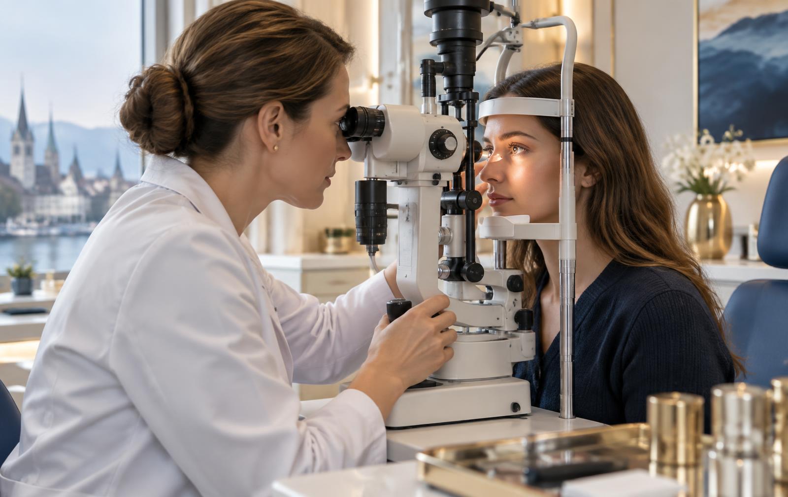
With every complete blink, the lids express a small amount of meibum from the gland openings along the lid margin. This secretion forms the outermost layer of the tear film and markedly slows evaporation.
If this lipid layer is missing or altered in quality, the tear film breaks up too early between blinks. The result is burning, fluctuating vision and a surface that reacts to the slightest irritation.
How dysfunction develops
When the secretion thickens or the gland ducts narrow, the glands become obstructed. Over months and years, gland segments can be remodelled or lost – a process that cannot be fully reversed.
Contributing factors include infrequent and incomplete blinking, lid-margin inflammation, rosacea, hormonal influences and inadequate lid hygiene.
Meibography: making gland structure visible
Using infrared meibography on the Topcon TERA Dry Eye Imager, we visualise the gland structure of the everted lid without touching the eye. The image allows shortening, thinning and gland loss to be assessed and documented in a standardised way.
We additionally record NIBUT, tear meniscus height, lipid layer dynamics and blink quality. Only this complete picture supports a reliable classification.
What makes sense therapeutically in MGD
The foundation is consistent lid hygiene, warming applications and baseline therapy matched to the findings. Where this is not sufficient, light-based modalities such as OPE® IPL may be considered for suitable indications, designed to act on processes in the lid-margin area.
Because gland loss is permanent, one principle applies: the earlier MGD is detected, the more structure can be preserved.
This website provides general information and does not replace an individual ophthalmic examination, diagnosis or treatment. Outcomes vary between individuals. Seek urgent medical care for sudden pain, loss of vision or eye injury.
More articles
All articles
Fundamentals2 September 20266 min read
Dry Eye: Causes, Symptoms and What Actually Helps
Burning, grittiness, watery eyes: dry eye usually has several drivers at once. An overview of tear film, inflammation and environment.
Read the article
Everyday life5 August 20265 min read
Dry Eyes and Screen Work: 8 Measures for the Working Day
At a screen we blink far less often and less completely. Eight practical measures that stabilise the tear film during the working day.
Read the article