Dry Eye: Causes, Symptoms and What Actually Helps
Fundamentals2 September 20266 min read
Dry eye disease is one of the most common diagnoses in ophthalmology – and one of the most underestimated. Understanding its causes explains why eye drops alone are often not enough.
Medically reviewed by the ophthalmic team of Lux Augenzentrum, Zurich

What dry eye means medically
Dry eye disease is a disorder of the tear film and the ocular surface. The tear film consists of an aqueous phase, a protective lipid layer produced by the Meibomian glands, and a mucin layer that helps it adhere to the surface. If one of these components is out of balance, the surface becomes unstable.
Professional societies broadly distinguish evaporative dry eye, where tears evaporate too quickly, from reduced tear production. In practice, mixed forms are common – which is why a differentiated assessment matters.
Symptoms worth taking seriously
Burning, a gritty or foreign-body sensation, tired eyes in the evening, light sensitivity, fluctuating vision and – paradoxically – watery eyes are among the most frequent complaints. Red, crusted or tender lid margins are an equally important sign.
Symptoms persisting for weeks, worsening during screen work or changing your contact lens tolerance should be assessed by an ophthalmologist.
The most common causes at a glance
Meibomian gland dysfunction (MGD) is considered the most frequent driver of evaporative dry eye. Further contributors include eyelid-margin inflammation, a reduced blink rate during screen work, hormonal changes, medications such as antihistamines or beta blockers, autoimmune conditions, allergies and climate factors such as air conditioning, heated indoor air and dry alpine air.
Previous eye surgery, long-term contact lens wear and cosmetics applied to the lid margin can also place stress on the ocular surface.
Why eye drops alone are often not enough
Tear substitutes reliably relieve symptoms and remain an important part of baseline care. They do not, however, remove the cause: if the Meibomian glands are obstructed or the lid margins inflamed, the tear film becomes unstable again as soon as the drops evaporate.
This is why we first measure objectively which component is affected – using meibography, non-invasive break-up time (NIBUT), tear meniscus height, lipid layer assessment and blink analysis.
How a personalised treatment plan takes shape
Consultation, clinical findings and measurements combine into a staged concept: baseline therapy and lid hygiene, anti-inflammatory measures where indicated and – where clinically appropriate – light-based modalities such as OPE® IPL and LLLT® photobiomodulation with eye-light®.
Whether light therapy is an option for you is always decided by ophthalmic assessment. Outcomes vary between individuals and are reviewed over time.
This website provides general information and does not replace an individual ophthalmic examination, diagnosis or treatment. Outcomes vary between individuals. Seek urgent medical care for sudden pain, loss of vision or eye injury.
More articles
All articles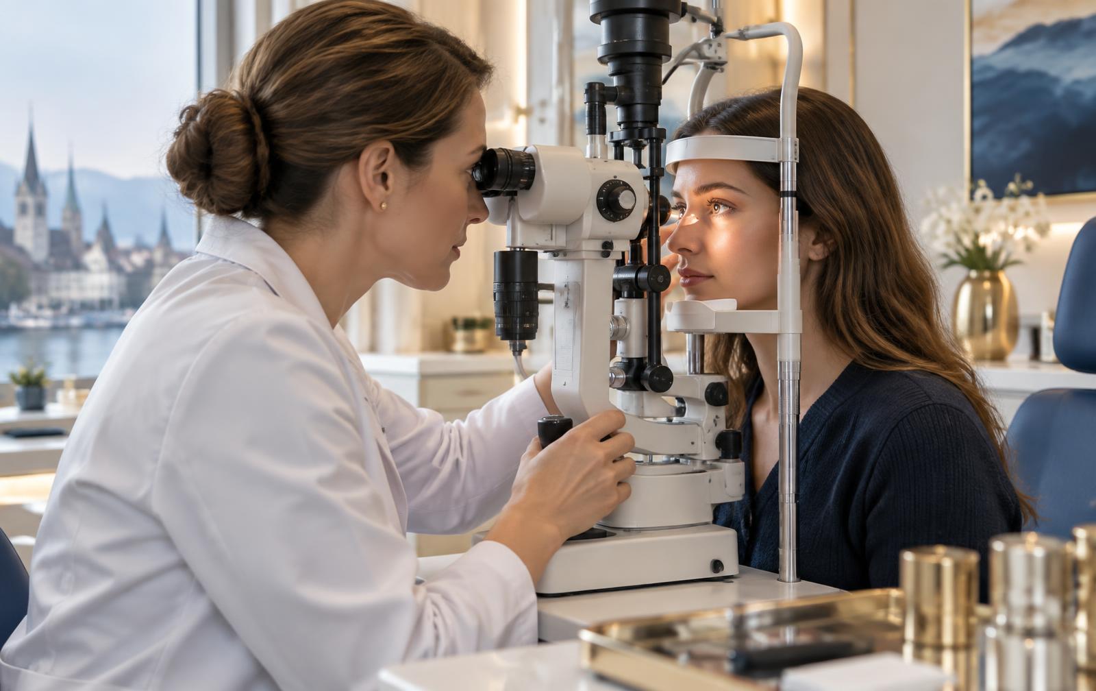
Diagnosis19 August 20265 min read
Meibomian Gland Dysfunction (MGD): The Underestimated Main Cause
The Meibomian glands supply the lipid layer of the tear film. How dysfunction develops – and how it can be documented objectively.
Read the article
Everyday life5 August 20265 min read
Dry Eyes and Screen Work: 8 Measures for the Working Day
At a screen we blink far less often and less completely. Eight practical measures that stabilise the tear film during the working day.
Read the article